(source: Electronics World, Aug. 1963)

New techniques and instruments now under construction may allow researchers to actually see the atomic structure of matter for the first time. Description of the transmission microscope, electron- and ion-emission types, proton microscope, and ultraviolet type.
By KEN GILMORE
SOMETIME during this year, a dramatically improved electron microscope may open up to scientists a completely new view of the universe.
Where current electron microscopes reveal for the most part only molecules, these new devices may allow researchers to actually see the atomic structure of matter--a long-time dream of scientists.
If the new instrument, now under construction at the University of Arizona, works as well as preliminary experiments indicate it will, it could have far-reaching effects on electronics, metallurgy, medicine, and other fields of science. But perhaps its most important impact Will be in biology, where scientists are now on the threshold of decoding the complex DNA molecule, the substance which may hold the key to the mystery of Life itself.
The instrument that promises to play a starring role in this coming scientific triumph is the latest in a family of electronic magnifying devices stretching back to the early part of the century. Included among its ancestors and current relatives are several generations of electron microscopes as well as a group of such related devices as the electron-emission microscope, ion-emission microscope, the proton microscope, and the ultraviolet flying-spot scanner.
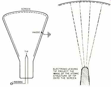
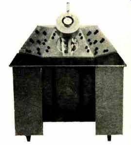
Fig. 1. The field-emission microscope is closely related to the cathode-ray
tube. Electrons are stripped by a high voltage from the tip of a needle
made of a metal whose structure is to be investigated. These electrons are
fired at a screen.
Magnification depends on ratio between tip and screen sizes.
Need for Electronics
The marriage of microscopy and electronics actually had its origin in a discovery in the 19th century, although no one knew it at the time. Ernest Abbe, a British scientist, published an analysis in 1875 showing that no object could be seen if it were much smaller than the wavelength of the light used to illuminate it.
This meant that nothing smaller than about 2000 angstroms* (approximately 1/125,000th of an inch) could ever be seen with a light microscope. Since microscopes of the day didn't have lenses good enough to approach 2000 angstroms resolution, the point was academic.
[*One angstrom unit equals 3.937 x 10^-9 inch or one hundred millionth of a centimeter . ]
But, in time, lenses improved. With a resolution of 2000 angstroms, the best instruments could reveal a few large molecules and many bacteria, microscopic plants, and other fascinating particles never before seen by man. But shall molecules, viruses ( whose existence was not even suspected at the time), and atoms were far beyond the instrument's power.
One early attempt to get around the fundamental law of nature which limits resolution took place in the early years of the 20th century when J. E. Barnard, an English microscopy expert, developed an instrument using ultraviolet, rather than visible light. The image couldn't be seen by the eye, since ultraviolet light is invisible, so it was focused onto a photographic plate which is sensitive to ultraviolet.
The ultraviolet microscope offered improved resolution, twice as good as a visible light microscope, but its greatest magnification was still far less than researchers wanted.
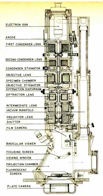
Fig. 2. Cross section of Norelco EM-200 electron microscope.
The first real step toward major improvement in microscopy came in 1924. Louis de Broglie, the distinguished French nuclear physics pioneer, speculated that elementary particles, especially electrons, in some ways demonstrate a wave-like nature similar to light. He derived the formula: wavelength = h /mc, where h is Planck's constant; m, is the mass of the particle involved; and c is its velocity. If an electron were accelerated to a velocity produced by an accelerating field of 60,000 volts, its wavelength accordingly would work out to be close to one-twentieth of an angstrom--about 1 /40,000th of a wavelength of light. If an instrument using electrons, instead of light, could be built, its theoretical resolution would then be 40,000 times better than that of the finest light microscopes in existence.
Early Instruments
During the next fourteen years, scores of instruments were built, and many worked, to a degree. First to operate were the emission microscopes ( Fig. 1) . A stream of electrons from the cathode was magnified as it passed through a series of electrostatic or electromagnetic lenses ( not shown ) , then projected onto a fluorescent screen. It gave a tremendously enlarged picture of the cathode itself, since the electrons from the cathode projected the cathode's microscopic structure onto the screen. The emission instrument, the first true electron microscope, was bypassed because the only image it can magnify is the image of the cathode itself. But recently, both emission and ultraviolet microscopes have been the subject of renewed interest, since they still offer certain unique advantages.
Although the work with emission instruments was encouraging, most early efforts went into the building of transmission microscopes--the kind generally used today. Here the electron stream passes through a thin specimen and projects the picture of that specimen on the screen. Almost anything of interest to science can be viewed with such an instrument.
Several working transmission instruments were built in the mid1930's. Then, in 1938, came a milestone in the history of microscopy. H.O. Müller and E. Driest, working in Germany, completed the first electron microscope whose resolution exceeded that of the best light instruments.
Development from that point was rapid. Less than a year later came an instrument that could resolve 100 angstroms and see particles 20 times smaller than anything viewed before. By 1940, resolution had improved so that objects only 20 angstroms across could be seen and RCA put the first commercial instrument on the market. By 1945--with dozens of the devices now making vast contributions to fundamental sciences in laboratories around the world-resolution reached about 10 angstroms. There, improvement all but stopped and even today the best instruments resolve no more than approximately 6-8 angstroms under optimum conditions.
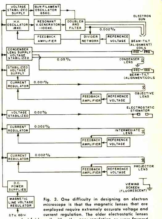
Fig. 3. One difficulty in designing an electron microscope is that the
magnetic lenses that are employed require extremely accurate voltage and
current regulation. The older electrostatic lenses worked fairly well with
rather poor regulation, so were favored many years ago when voltage-regulation
techniques were poorer. The inherently greater distortion of these designs,
though, has made their use in microscopes quite rare today.
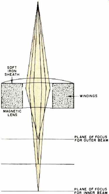
Fig. 4. Parts of the electron beam passing through a magnetic lens near
the center do not come into focus at the same plane as those passing through
nearer the outside of the electron beam. This phenomenon is known as spherical
aberration.
Operating Principles
The modern electron microscope (see cover and Fig. 2) is actually nothing more than a large, highly specialized cathode-ray tube. An electron gun at the top of the column generates a cloud of electrons which are attracted by the anode a short distance away. This electrode, normally operated at 50,000100,000 volts positive with respect to the cathode, attracts the electrons strongly. They move toward the anode.
( Actually, the anode and the rest of the instrument are usually at ground potential for safety's sake, and the insulated electron gun is held at a high negative potential.) The electrons accelerate to a high speed, shoot through the hole in the anode, and continue on down the column to the viewing screen and photographic plate at the bottom.
They pass through a magnetic field ( generated by the condenser lens, actually a coil) shortly after leaving the anode, and are focused into a tight beam as they hit the specimen. A section of the beam hitting the dense section of the specimen is largely absorbed, another part hitting a thinner area passes through with little or no attenuation.
The beam, retaining the varying pattern of electron density impressed on it as it went through the specimen, then goes through a series of magnetic objective and projector lenses (coils) which magnify it. Finally, it reaches the bottom of the column where it falls on a fluorescent screen and reproduces--much magnified--the image of the specimen. A camera is arranged either to photograph the luminous screen, or else to allow the electrons themselves to fall directly on an electron-sensitive photographic plate.
Construction is Critical
Although the electron microscope is simple in principle, its mechanical and electronic design (Fig. 3) present severe problems. Most of them--especially those which limit resolution--are associated with the electronic lens.
In a modern magnetic lens (electrostatic lenses, once popular, are rarely used today because of their greater inherent distortion) the magnetic lines of force are confined by a soft-iron-sheathed coil. The electron beam, shooting through this field, spreads into a cone shape and is greatly magnified, like a light ray passing through a glass lens.
The trouble comes in meeting critical construction tolerances. Pole pieces machined to the highest standards consistent with current production techniques and the most accurate lens coils available are not good enough. Minor, but unavoidable, imperfections and traces of contamination put kinks in the magnetic field big enough to distort the electron beam.
For this reason, compensating circuits are included and final alignment is done by watching the image produced while adjusting the instrument for least distortion.
Another problem is spherical aberration. Electrons passing through the center of the magnetic lens converge (come into focus) at one distance, those passing through the lens away from the center, at another (Fig. 4). If the part of the picture represented by the center of the beam is in focus, other parts are hazy.
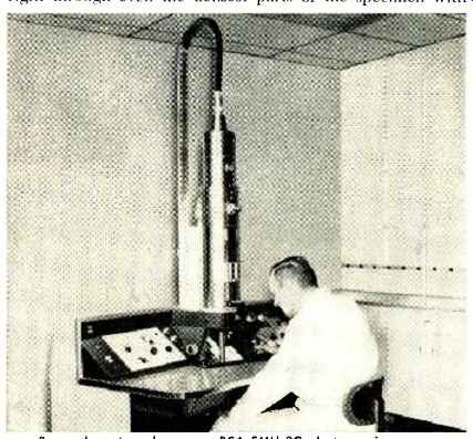
---Researcher at work on new RCA EMU-3G electron microscope.
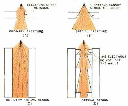
Fig. 5. (A) Configuration of conventional and (B) improved lens structure.
The lens is cone-shaped, with the angle of the cone's inner surface being
greater than the angular divergence of the electron beam. The beam then "sees" only
the thin lip of the lens and is not affected by imperfections on most
of the inner surface. (C) and (D) show how this new technique has been
applied to the design of a complete column.

Fig. 6. Cross section of microscope designed by Dr. Wilska.
One reason electron microscopes are operated at high voltages is that high-velocity electrons, like a speeding bicycle rider, are more stable than slow-moving (low-voltage) ones.
But this high-voltage technique--used almost exclusively on modern instruments--brings its own problems.
The most serious one is that fast-moving electrons have relatively greater penetrating power. They are likely to whiz right through even the densest parts of the specimen with few being stopped, and no image being formed. Consequently, microscopists use relatively thick specimens, which contain enough matter to stop part of the electron beam. In practice, the thinnest practical specimen is about 200 angstroms. And, as a general rule, it is impossible to see specimen details in an electron photomicrograph smaller than about 1 /10th the thickness of the sample. With a 200angstromthick specimen, maximum resolution is about 20 angstroms.
In some cases it can be reduced to half that and, in exceptional cases, objects 6 to 8 angstroms have been resolved.
This puts scientists tantalizingly close to one of their major goals--to see the atoms in biological molecules of greatest interest, such as DNA. These atoms, it is estimated, are some 2 to 3 angstroms in diameter. This is within the theoretical resolving power of the electron microscope, but it has not as yet been achieved.
Promising New Developments
The work of Dr. Alvar Wilska, a Finnish-born scientist now at the University of Arizona, is attracting attention because his two major improvements in the electron microscope promise to extend its resolving power into this vital region. To get around the thickness problem, Dr. Wilska lowered the accelerating voltage so that the electrons could be stopped by thinner sections. But slower-moving electrons are far more sensitive to lens imperfections, electrostatic charges on the column, and stray magnetic fields.
Dr. Wilska built a series of low-voltage instruments in which he gradually improved the lens design to the point where he got usable pictures with accelerating voltages as low as 1000 volts. His main design innovation was to build a special cone-shaped lens so that the electron beam would "see" less of the lens as it passes through, and therefore be less affected by its imperfections (Figs. 5 and 6). This improvement gave high-contrast pictures from specimens only a few angstroms thick and thus eliminated one fundamental limitation to better resolution. But the resolving power of the microscope was improved little, if any, because of the effects of spherical aberration.
Several years ago, Wilska set to work on this problem.
Since spherical aberration is caused by electrons passing through various parts of the lens and coming into focus at different planes, the solution was to make them all go through the lens at the same distance from its axis.
Wilska blocked the center of the lens (Fig. 7A) so that all electrons must pass through a narrow annular slit. An experimental lens built on this principle worked as predicted.
A full-scale microscope, using both the low-voltage and spherical–aberration-correcting techniques worked out by Wilska is now under construction. Within the year, it may let man see the atoms of which living matter is made.
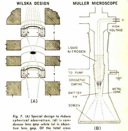
Fig. 7. (A) Special design to reduce spherical aberration. (d) is condenser
lens gap while (e) is objective lens gap. Of the total cross section of
the electron beam, only a narrow annular portion passes between marginal
ring (a) and central stopper (b). Both (al and (b) are at the same positive
potential as the column itself, while there is an electrostatic field between
negatively charged ring electrode (c) and the central stopper (b). This
field increases in strength toward the center thus counteracting effects
of spherical aberration within the zone between (a) and (b). (B) Dr. Miller's
ion emission microscope achieves greater resolution than its predecessor,
the field emission microscope, by using ions instead of electrons to fire
at the screen and reproduce an image of the atomic lattice of the emitting
tip. If the tip is cooled enough, by use of liquid nitrogen, helium molecules
will stick long enough to lose kinetic energy. Thus, these molecules become
much easier to ionize.
Other Approaches
As de Broglie's formula shows, mass of the illuminating particle is inversely proportional to wavelength. Therefore, greater theoretical resolution could be achieved by using particles heavier than electrons. Such particles might also be more stable and less subject to distortion by lens contamination and stray magnetic fields. Putting these principles to work, a team of French scientists at the National Research Center in Paris has built a microscope which uses protons instead of electrons. Although this approach has attractive possibilities, there are many practical problems. So far, no proton or other heavy-particle instrument has achieved the resolution possible with electron microscopes.
Meanwhile, as the transmission electron microscope held the center of attention of both the scientific community and the world at large, other earlier devices, bypassed in the search for more widely useful instruments, have not been allowed to languish completely. For example, a number of scientists, including Dr. Erwin Miiller of Pennsylvania State University, have continued to work on emission microscopes. As early as 1936, Willer demonstrated an instrument of this type with a resolving power of 20 angstroms, far better than results achievable with transmission microscopes of the day. Its resemblance to an ordinary cathode-ray tube is striking.
--Norelco EM-200 electron microscope.
The maximum resolution of Müller's instrument was limited by the fact that electrons, being very light, are apt to be easily diverted by stray magnetic fields or other conditions. So Müller decided to use a heavier particle. Several years ago, his first operating model of the ion-emission microscope (using ions instead of electrons) was demonstrated. (The ion, being heavier, also has a shorter wavelength and is thus inherently capable of better resolution than the electron.) Just a short time ago, the first commercial model, made by Cenco Instruments, came on the market.
The ion microscope ( Fig. 7B) is quite similar to the electron-emission instrument. A small amount of helium gas is introduced into the evacuated tube. As an atom of helium floats to within a few angstroms of the tip, the high voltage there tears an electron away from it. The helium--now a positively charged ion with an electron missing--is repelled violently from the tip of the needle toward the screen. The patterns of ions hitting the screen is a faithful reproduction of the atomic lattice at the end of the needle.
With this arrangement, Müller has succeeded in getting magnification up to 10,000,000 and resolution of less than 3 angstroms. At 10,000,000 the picture is very dim, so 2,000,000 is the approximate maximum practical amount of magnification.
The ion-emission microscope has one serious drawback; it can be used only to examine metals, and then only a handful which have melting points above 1400°C. Metals which melt at a lower temperature vaporize when subjected to the high voltage necessary to ionize the helium atoms, and therefore cannot be used.
In spite of its limited applications, Müller's ion microscope is becoming increasingly important. Scientists are now using it to find out what really happens when a wire is annealed, to learn more about the crystalline structure of metals, to understand metal fatigue. Soon, we may approach a broader understanding of the mysterious action of catalysts.
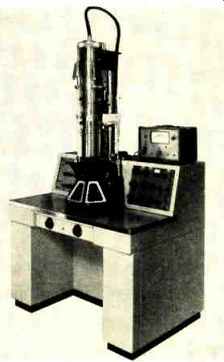
-------Experimental model of Wilska's short-column low-voltage unit.

----Newly commercialized version of Müller field ion microscope.
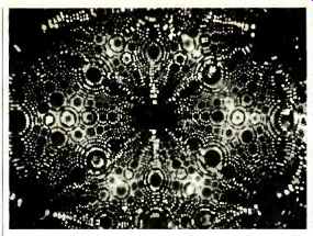
----- Crystalline structure of tungsten is shown on photomicrograph
taken with field ion microscope. Each luminous spot in the photomicrograph
represents an individual atom of matter.
Another offshoot from the early days of microscopy is the flying-spot ultra-violet microscope. The original ultra- violet instrument had several important defects which kept it from becoming widely used back in the early years of the century. One of the most important was that ultraviolet light quickly kills most forms of microscopic life which scientists were interested in observing.
(It shared that fault with the electron microscope, of course.) But two researchers at the University of Texas came up with a variation a few years ago which overcame this problem.
They generated a spot on the face of an ultraviolet cathode-ray tube, which they swept in a raster through an ultraviolet microscope onto a specimen they wanted to study. The spot scans the specimen just as the beam in a TV camera scans the scene before it. Under the specimen, a photomultiplier tube collects the light, which varies in intensity as the transparency of the specimen it is sweeping varies.
The fluctuating signal developed by the photomultiplier is used to modulate the beam of a television picture tube, which is sweeping in synchronism with the ultraviolet spot. Since the spot is small and sweeps rapidly across the specimen, it has no effect on the organism being studied.
Although the combination of electronics and microscopy has already produced results of incalculable value, it seems likely that its future contributions may be even more spectacular. RCA's Dr. V. K. Zworykin, a pioneer in the field, recently put his thoughts on the electron microscope and its importance this way: "Today, we know that the electron microscope has opened a great new dimension for human exploration in the world which we once labeled submicroscopic. The knowledge that has already resulted is surely only a small portion of that which has been brought within our ultimate reach as science continues to apply the electron microscope to the endless task of research."