
Fig. 1--The scopes used by the medical profession are not the same as
service scopes. An experienced biomedical electronic technician tells
how medical scopes are different.
Most medical-electronics wave forms are viewed on oscilloscope (scope) cathode-ray tubes (CRTs). When permanent traces are needed, similar waveforms are recorded on moving tape of strip-chart recorders (oscillographs). But the trouble and expense of the recorders discourage other uses. Strip recorders are far outnumbered by scopes.
These scopes range between large multichannel models (that allow the simultaneous monitoring of as many as eight patients, as shown in Figure 1) to small single-channel types. This covers a variety of features and circuits.
How are medical scopes different? Medical scopes are not the same as electronic-service scopes. The major differences and similarities follow.
Sweep speeds--Medical scopes are used to display waveforms produced by physiological functions. Most of these change slowly. The human heart, for example, normally operates at 40 beats per minute (BPM) to 130 BPM. That's between 0.66 and 2.2 "cycles" per second. So, medical scopes must have very slow horizontal-sweep rates.
Typical sweep speeds are 25 mm/s, 50 mm/s, arid 100 mm/s.
The standard for electrocardiograph display is 25 mm/s. On a conventional CRT screen of 10 cm, this translates to one sweep every four seconds. A heartbeat of 60 BPM produces four complete wave forms across the entire screen at all times. (According to triggered-sweep ratings, that's a sweep time of 0.4 S/div. For recurrent-sweep scopes, it corresponds to a sweep frequency of 1/4 Hz. Incidentally, the formula 1/4 Hz = 4s, doesn't make much sense until Hertz is translated to its literal meaning. Then it reads 1/4 cycles/s = 4 s/cycle.) Sensitivity-Several sensitivities of vertical gain are found in medical scopes. However, each scope model usually has only one. If the scope is used as a display device for other instruments, the sensitivity typically will be between 0.5 V/cm to 2 V/cm. Scopes used for direct EEG or ECG monitoring (without other instruments) have sensitivities of a few millivolts or microvolts per centimeter.
Phosphor specs--One easily-apparent difference between TV-service scopes and medical scopes is the color and persistence of the phosphor inside the CRT screen.
Service and lab scopes must display fast-rise-time or varying-amplitude waveforms that are traced at rapid sweep speeds. So, the phosphors must have short persistence. That is, the brightness must fade rapidly after the writing beam has moved away.
If medical low-frequency wave forms and sweep speeds are displayed on a CRT . having short persistence, nothing can be seen except a single bright dot that leisurely moves up and down as it proceeds slowly from left to right.
No waveform details can be perceived under those conditions.
The brightness of a long-persistence (P7) phosphor in a medical scope fades slowly so the waveform can be seen as a long "comet tail" following the moving spot. When the beam reaches the right side, the left edge of the trace is almost extinguished. But it can be seen well enough to allow a satisfactory diagnosis of most waveform irregularities.
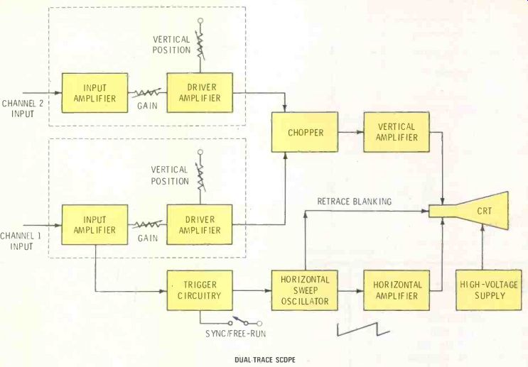
Figure 2--The block diagram of a service scope and a "bouncing-ball" medical
scope are just alike. There are major differences of sweep speed, bandwidth
and phosphor persistence.
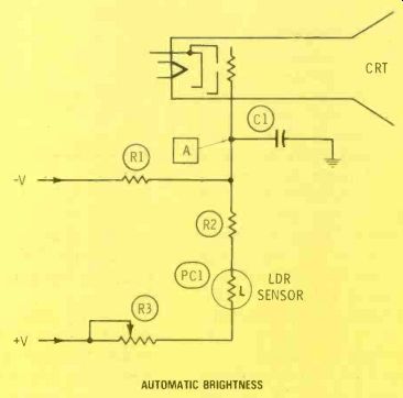
Figure 3--Ambient light falling on a photocell adjusts the brightness
of many scope models by varying the CRT grid bias.
Incidentally, the phosphors chosen for medical scopes glow with large amounts of yellow and blue-violet tints. Some manufacturers add filters that reduce the yellow, thus the trace appears to be violet.
Others filter out the violets and blues leaving a bright yellow wave form. However, the same CRT might be used in scopes showing either tint. Don't be surprised to see a blue or violet color from the rear of the CRT, but a yellow trace through the filter in front! "Bouncing-ball" displays Non-memory types of medical scopes usually are called "bouncing-ball" models, because of the moving spot .with the fading trace that follows it. The majority of older scopes are of this type.
Figure 2 shows the block diagram of a bouncing-ball medical scope.
This specific scope is a 2-channel model. However, single-channel versions are essentially the same, except for the chopper and the extra vertical amplifier.
Probably you will notice no distinctive differences between service and medical scopes from their respective block diagrams. All medical scopes have triggered sweep.
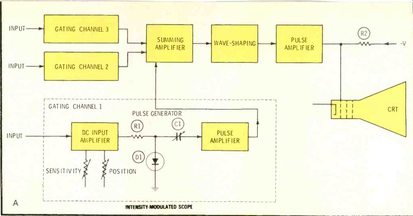
Figure 4 (A) Waveforms of several channels in San born scopes are produced
by intensity modulation of the CRT brightness while the entire raster
is scanned constantly. Phase of sampling pulses determines the position
of each wave form, as shown by the (B) drawing.
Deflection--Because of the wide bandwidth signals handled by service-type scopes, they have electrostatic deflection. Some medical waveforms can be displayed adequately by flat response only to 200 Hz, and none requires more than 3000 Hz. Such narrow bandwidths are well within the ability of magnetic yokes.
Therefore, the majority of medical scopes are deflected magnetically, since equal performance can be obtained in a shorter CRT.
Automatic brightness
Some medical scopes include a photocell that adjusts the CRT brightness according to the amount of light in the room. An intensity setting that's appropriate with both bright fluorescent lights and some daylight would prove an annoying distraction at night when the room is darkened so the patient can sleep. Therefore, these automatic brightness features are very practical, and not an unneeded luxury.
An example of automatic brightness is illustrated by the schematic in Figure 3. A photoresistor controls the grid bias of the CRT.
Grid bias of the CRT is produced by a delicate balance of voltages from positive and negative sources, so the grid has the proper negative voltage relative to its cathode.
When the ambient light reaching the photocell is strong, the photo cell resistance increases the amount of positive voltage fed to the grid circuit, and the negative grid bias is decreased. Reduced negative grid bias allows the CRT to draw more gun current and produce a brighter trace.
Of course, when the room is darkened, photocell PC1 resistance increases, and less positive voltage is sent to the grid circuit. In turn, less negative voltage is cancelled, which increases the CRT negative bias and decreases the waveform brightness.
Multichannel scopes
A conventional chopper electronic switch allows the scope of Figure 2 to display two waveforms on a conventional CRT. A high-repetition-rate square wave alternately connects one vertical signal and then the other to the vertical amplifier. This is sampling of the waveform, which reconstructs each waveshape out of many short lines.
These lines are not very visible because of the long persistence of the phosphor and the lack of synchronism between signal and chopper frequencies.
More than two channels can be accommodated by adding another electronic switch and increasing the switching frequency. Also, there is a different method, which is de scribed next.
Pulse-timing operation--Another way of obtaining several vertical channels in a scope that's equipped with a conventional CRT is to use the pulse-timing circuit of Figure 4.
Originally, the design was pioneered by Sanborn (now it's manufactured by Hewlett-Packard).
A 3-channel scope with pulse timing circuits is shown in the block diagram of Figure 4A. Details are shown only for one channel, but the other two are identical.
Because the waveforms are not produced by varying the amount of deflection (as it's done in service scopes), the raster is scanned by both vertical and horizontal sweeps at all times. The waveforms are formed by intensity modulation which permits CRT current only at proper times.
This kind of raster scanning can be compared to deflection in a TV receiver. However, there are important differences.
About three seconds are required for the horizontal sweep to move the beam completely across the screen. That much is similar to the operation of other medical scopes.
The vertical sweep works some what like the horizontal sweep does in TVs (except the beam is moved vertically rather than horizontally).
In fact, the Sanborn version uses a 6CD6/6CB5 TV horizontal-output tube as a vertical-output tube that drives a regular flyback transformer (replaceable by a Triad D-604).
Frequency of the vertical sweep is not critical, since it is not locked or synchronized, but usually it is adjusted above 15 kHz to minimize squeals that some people find annoying.
Each vertical channel has a dc amplifier, a tunnel-diode pulse generator and a pulse amplifier. Pulses from all channels are summed in the main channel, and after shaping are applied to the CRT control grid.
Timing of each pulse generator depends on the instantaneous signal that's applied to the pulse genera tor. This signal is made up of the input analog signal (after amplification) plus a dc voltage from the positioning control. With zero voltage applied, the time between pulses will occur at a constant (resting) rate. A positive or a negative voltage causes the pulses to be produced faster or slower.
The CRT is biased to cut-off until a pulse reaches the grid.
Consequently, pulses that arrive at an earlier time make dots of light nearer the top of the screen, while the slower-arriving pulses cause the lighted dots to be seen lower on the CRT screen.
An analog signal affects this channel line in the same way, except the voltage is varying. There fore, the pulses arrive earlier or later, moving the line of dots up or down to form the waveshape.
Figure 4B shows horizontal lines of dots for channels 1 and 2 because they have no analog input signal. Channel 3 dots trace the shape of the analog input signal. Of course, the number of vertical lines in the drawing has been minimized to make the operation clear. Actually, there are about 45,000 vertical lines across the total screen. So, the lines appear to be continuous.
Most repairs to this type of scope will be in the vertical-sweep system.
Bad damper or output tubes, shorted flyback transformers and oscillator problems cause most of the service work.
Figure 5 This digital non-fade scope operates ECG, arterial pressure and patient temperature, on batteries while displaying

------ DIGITAL NON-FADE SCOPE
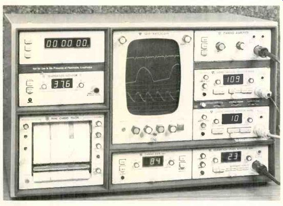
------ One rack holds a strip-chart recorder, two blood-pressure
monitors, cardio amplifier, respiration monitor, temperature module,
cardio-rate module, a timer and a four-channel scope. Similar monitors
are used in intensive-care units (ICU) and operating rooms.
Non-fade models
Even long-persistence phosphors can't eliminate fading of the left part of a waveform as the beam reaches the right edge. This creates problems for doctors because many small details of the waveform are transient and sometimes not repeated for some time. Therefore, bouncing-ball scopes force them to guess about some waveform anomalies.
The problems are solved by a special non-fade (digital) type of medical scope that uses an analog-to-digital (A/D) converter to change the analog signal voltages into digital equivalents which can be stored in various kinds of memories. When needed for display, a memory signal is decoded by a digital-to-analog (D/A) converter.
The resulting analog signal is amplified and viewed on the CRT.
With constant digital refreshing, the real-time waveforms move across the screen with full brightness. Stored non-refreshed digital signals can freeze a waveform for detailed study.
Keep in mind that these non-fade scopes are not like the storage scopes used in laboratories and for research. Storage scopes use a CRT of special design which holds the last trace on the screen for a time.
A portable ECG monitor manufactured by American Optical is shown in Figure 5. This non-fade scope displays ECG, arterial pressure and patient temperature while operating from internal batteries.
Notice the button labeled FREEZE. It holds the current display on the screen even after the input signal has been disconnected.
Digital scope circuits--Figure 6 shows the block diagram of a typical single-channel non-fade scope, and the block diagram of a 4-channel scope is given in Figure 7.
In both scopes, the analog input signals first must be digitized in an A/D converter. Consider an example of an 8-bit A/D converter which can represent 28-1 (255) different levels of amplitude. Therefore, it's convenient for the A/D converter to have a full-scale capacity of +2.55 V, so the decimal equivalent of the binary word is numerically the same as the voltage it represents. In binary, full scale is 11111111 (or +2.55 V) half scale is 10000000 (+1.28 V) and zero is 00000000.
Output of the A/D converter is stored in two stages of a random access memory (RAM) integrated circuit (the same type as used in microcomputers). A small "scratch-pad" memory holds four successive 8-bit words, while most of the data is held in a 1024-word main memory.
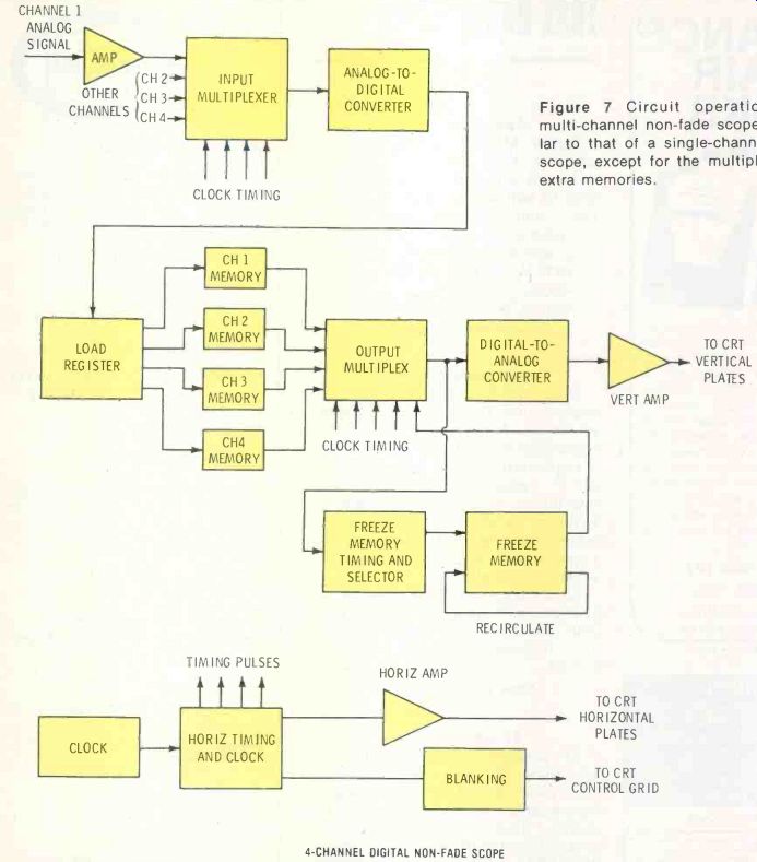
Figure 7---Circuit operation of a multi-channel non-fade scope is similar
to that of a single-channel digital scope, except for the multiplexer
and extra memories.
Data in the scratchpad memory is the most recent, and for practical purposes represents the real-time signal amplitude. A scope deflection system responds only to analog voltages or currents, so a digital-to-analog converter is provided to decode the memory output and supply the analog signal to the vertical scope amplifiers.
A multi-phase-system clock synchronizes the action of this circuit.
It generates an ADC START pulse to initiate one A/D conversion, and the results are stored in the next sequential location of the scratch-pad memory.
A memory read/write line commands the memory either to write data into or read data out of the memory location addressed by the address lines. The memory is scanned by incrementing the ad dress lines with clock pulses (a binary counter is used as an address generator). These same clock pulses are used to control the horizontal sweep.
The refresh rate usually is around 64 times per second. During refresh, the W/R line will be in READ mode, and the memory is scanned by successively incrementing the address generator. Each successive memory location contains an 8-bit digital word which represents an amplitude point on the input waveform, so the digital-to-analog converter output will follow the original waveshape.
At the beginning of each scan, contents of the scratchpad memory are loaded into the first four locations in the main memory. This provides a continual update of the data, and makes the display appear to sweep like a regular scope.
A multiplexed 4-channel non-fade scope is shown in the block diagram of Figure 7. It is essentially similar to the previous example, except for needing four memories to accommodate the four channels, and requiring both analog and digital multiplexers.
The memory-freeze circuitry is capable of transferring the contents of any one of the channel memories to a separate memory bank that is not updated by the input signal data. This memory has its output fed back to its input, so it will recirculate the same data continuously without any updating. The effect is to provide a display that's frozen on one waveform. The freeze memory is viewed on a separate multiplex channel, so its trace will appear at the bottom of the screen. Thus, it's possible to see the real-time trace and a frozen segment at the same time for wave form comparisons.
Comments
This general information should have clarified the important differences between service and medical scopes, and provided sufficient facts to help you service them.
Also see: Tips for using scopes--part 1